カテプシン
| Cathepsin | |||||||||
|---|---|---|---|---|---|---|---|---|---|
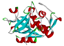 カテプシンKの構造 | |||||||||
| 識別子 | |||||||||
| 略号 | CTP | ||||||||
| Pfam | PF00112 | ||||||||
| Pfam clan | CL0125 | ||||||||
| InterPro | IPR000668 | ||||||||
| SMART | Pept_C1 | ||||||||
| PROSITE | PDOC00126 | ||||||||
| MEROPS | C1 | ||||||||
| SCOP | 1aec | ||||||||
| SUPERFAMILY | 1aec | ||||||||
| |||||||||
| テンプレートを表示 | |||||||||
カテプシン(英: cathepsin)は、全ての動物やその他の生物にみられるプロテアーゼである。カテプシンファミリーには10種類以上のメンバーが存在し、その多くはリソソームに存在して低pHで活性化される。こうしたメンバーの活性はほぼ完全にオルガネラ内に限られる一方で、カテプシンKのように細胞外で機能するものも存在し、このタンパク質は破骨細胞によって分泌されて骨吸収(英語版)過程で機能する。カテプシンは哺乳類細胞のターンオーバーに重要な役割を果たしている。
メンバー
- カテプシンA(英語版)(セリンプロテアーゼ)
- カテプシンB(英語版)(システインプロテアーゼ)
- カテプシンC(英語版)(システインプロテアーゼ)
- カテプシンD(英語版)(アスパラギン酸プロテアーゼ)
- カテプシンE(アスパラギン酸プロテアーゼ)
- カテプシンF(英語版)(システインプロテアーゼ)
- カテプシンG(英語版)(セリンプロテアーゼ)
- カテプシンH(システインプロテアーゼ)
- カテプシンK(システインプロテアーゼ)
- カテプシンL1(英語版)(システインプロテアーゼ)
- カテプシンO(英語版)(システインプロテアーゼ)
- カテプシンS(英語版)(システインプロテアーゼ)
- カテプシンV(英語版)(カテプシンL2、U、システインプロテアーゼ)
- カテプシンW(システインプロテアーゼ)
- カテプシンZ(英語版)(カテプシンZ、P、システインプロテアーゼ)
臨床的意義
カテプシンは、疾患への関与が示唆されている生理過程に多く関与している。システインプロテアーゼ型のカテプシンは、薬剤標的として多くの研究が行われている[1][2]。
- がん - カテプシンDは分裂促進因子であり、またケモカインの分解によって樹状細胞の機能を阻害することで腫瘍免疫応答を減弱させる。カテプシンB、Lは細胞外マトリックスの分解と細胞の浸潤に関与している[3]。
- 脳卒中[4]
- 外傷性脳損傷[5]
- アルツハイマー病[6]
- 関節炎[7]
- エボラ出血熱 - カテプシンB(そして程度は低いもののカテプシンL)はウイルスが宿主細胞へ侵入するために必要であることが知られている[8]。
- COPD
- 慢性歯周炎(英語版)
- 膵炎
- いくつかの眼疾患: 円錐角膜、網膜剥離、加齢黄斑変性、緑内障[9]
カテプシンA
カテプシンA(英語版)の欠乏はガラクトシアリドーシス(英語版)と関係している[10]。転移性メラノーマの病変部位では、原発巣と比較してカテプシンAの活性が有意に上昇している[11]。また、筋ジストロフィーや除神経が生じる疾患の影響を受けた筋肉では、カテプシンAが増加している[12]。
カテプシンB
カテプシンB(英語版)はβ-セクレターゼ1と同様に機能し、アミロイドβ前駆体タンパク質を切断してアミロイドβを生成している可能性がある[13]。カテプシンBはペプチダーゼC1ファミリーに属し、その過剰発現は食道腺癌やその他の腫瘍と関連している[14]。また、カテプシンBは卵巣がんなどの腫瘍のプログレッションへの関与も示唆されている[3]。
カテプシンD
カテプシンD(英語版)は、フィブロネクチンやラミニンなどさまざまな基質を切断しているようである。他の一部のカテプシンとは異なり、カテプシンDは中性のpHでもある程度のプロテアーゼ活性を有している[15]。腫瘍細胞におけるこの酵素の高発現は、浸潤性の高さと関連しているようである。
カテプシンK
カテプシンKは、哺乳類において最も強力なコラゲナーゼ(英語版)である。カテプシンKは骨粗鬆症に関与しており、この疾患では骨密度が低下し骨折のリスクが高まる。破骨細胞は体内で骨吸収を行う細胞であり、骨基質におけるミネラル以外の主要なタンパク質構成要素であるコラーゲンを分解するためにカテプシンKを分泌する[16]。また、カテプシンKは他のカテプシンとともに、細胞外マトリックスの分解によってがんの転移にも関与している[17]。カテプシンKをノックアウトしたアテローム性動脈硬化マウスは、アテローム病変のサイズが低下することも示されている[18]。培養血管内皮細胞におけるカテプシンKの発現はずり応力によって調節されている[19]。カテプシンKは関節炎に関与していることも示されている[20]。
カテプシンV
マウスのカテプシンLはヒトのカテプシンV(英語版)と相同である[21]。マウスカテプシンLはアディポジェネシス(英語版)や耐糖能障害に関与していることが示されている。カテプシンLはフィブロネクチン、インスリン受容体、IGF-1受容体を分解する。カテプシンL欠損マウスは野生型と比較して脂肪組織が少なく、血清グルコース濃度やインスリン濃度は低い。またインスリン受容体サブユニットや、グルコーストランスポーター(GLUT4)、フィブロネクチンは増加している[22]。
阻害剤
5種類の環状ペプチドが、ヒトのカテプシンL、B、H、Kに対する阻害活性が示されている[23]。カテプシンKやSを標的としたいくつかの阻害剤は骨粗鬆症、変形性関節症、慢性疼痛に対する治療薬としての臨床試験段階に到達しているが、カテプシンK阻害剤レラカチブ(Relacatib)、バリカチブ(Balicatib)、オダナカチブ(英語版)は、有害な副作用のためにそれぞれ第I相、第II相、第III相で試験が中止されている[24]。カテプシンS阻害剤SAR114137は、慢性疼痛を対象とした第I相試験を通過しなかった。2022年、カテプシンL阻害剤オルゴトレルビル(英語版)(STI-1558)はCOVID-19治療のための第I相試験開始のFDA認可を受けた[25]。
カテプシンザイモグラフィー
ザイモグラフィー(英語版)はゲル電気泳動の一種であり、酵素の基質を共重合したポリアクリルアミドゲルを用いて酵素活性を検出する手法である。カテプシンザイモグラフィーでは、ゼラチンを共重合したポリアクリルアミドゲル中の泳動度の差異によってさまざまなカテプシンの分離が行われる。電気泳動を非還元条件、ロイペプチン添加のもとで行うことで、酵素は不可逆的分解から保護される[26]。まずタンパク質濃度を決定した後、等量の組織タンパク質をゲルにロードし、電気泳動を行う。その後、ゲルを再生(renaturation)バッファーへ移すことで、カテプシンは天然状態のコンフォメーションへと戻される。そして、特定のpHの活性化バッファーへ移し、37 °Cで終夜インキュベーションする。この活性化段階では、カテプシンによるゼラチン基質の分解が行われる。このゲルをCBCを用いて染色すると、ゼラチンが残っている領域は青く染色されるのに対し、カテプシンの活性があった領域はゼラチンが分解されているため染色されず、白いバンドとして可視化される。カテプシンザイモグラフィーはfmol量の成熟カテプシンKを検出することができる[26]。さまざまなカテプシンはその分子量に応じた泳動度に基づいて同定される。また、各カテプシンにはそれぞれタンパク質分解活性が最大となる至適pHがある。一例としてカテプシンKはpH7または8でゼラチンを分解することができるが、カテプシンLやVはこのpHでは活性を示さない。逆にpH4ではカテプシンVは活性を有するが、カテプシンKは活性を示さない。そのため活性化バッファーのpHを調整することで、カテプシンの種類をさらに詳細に同定することができる[27]。
出典
- ^ Novinec, Marko; Lenarčič, Brigita (1 June 2013). “Papain-like peptidases: structure, function, and evolution”. BioMolecular Concepts 4 (3): 287–308. doi:10.1515/bmc-2012-0054. PMID 25436581.
- ^ Turk, Vito; Stoka, Veronika; Vasiljeva, Olga; Renko, Miha; Sun, Tao; Turk, Boris; Turk, Dušan (January 2012). “Cysteine cathepsins: From structure, function and regulation to new frontiers”. Biochimica et Biophysica Acta (BBA) - Proteins and Proteomics 1824 (1): 68–88. doi:10.1016/j.bbapap.2011.10.002. PMC 7105208. PMID 22024571. https://www.ncbi.nlm.nih.gov/pmc/articles/PMC7105208/.
- ^ a b “Involvement of cathepsins in the invasion, metastasis and proliferation of cancer cells”. J. Med. Invest. 52 (1–2): 1–9. (February 2005). doi:10.2152/jmi.52.1. PMID 15751268. http://medical.med.tokushima-u.ac.jp/jmi/vol52/pdf/v52_n1-2_p001.pdf.
- ^ Lipton P (October 1999). “Ischemic cell death in brain neurons”. Physiol. Rev. 79 (4): 1431–568. doi:10.1152/physrev.1999.79.4.1431. PMID 10508238.
- ^ “Inhibition of cathepsin S produces neuroprotective effects after traumatic brain injury in mice”. Mediators of Inflammation 2013 (2013): 187873. (Oct 24, 2013). doi:10.1155/2013/187873. PMC 3824312. PMID 24282339. https://www.ncbi.nlm.nih.gov/pmc/articles/PMC3824312/.
- ^ Yamashima T (2013). “Reconsider Alzheimer's disease by the 'calpain-cathepsin hypothesis'--a perspective review”. Progress in Neurology 105: 1–23. doi:10.1016/j.pneurobio.2013.02.004. PMID 23499711. https://zenodo.org/record/895448.
- ^ “Serine protease cathepsin G regulates adhesion-dependent neutrophil effector functions by modulating integrin clustering”. Immunity 22 (6): 679–91. (June 2005). doi:10.1016/j.immuni.2005.03.015. PMID 15963783.
- ^ Chandran K (2005). “Endosomal Proteolysis of the Ebola Virus Glycoprotein Is Necessary for Infection”. Science 308 (5728): 1643–1645. Bibcode: 2005Sci...308.1643C. doi:10.1126/science.1110656. PMC 4797943. PMID 15831716. https://www.ncbi.nlm.nih.gov/pmc/articles/PMC4797943/.
- ^ “The role of cathepsins in ocular physiology and pathology”. Exp. Eye Res. 84 (3): 383–8. (March 2007). doi:10.1016/j.exer.2006.05.017. PMID 16893541.
- ^ Kleijer, W. J.; Geilen, G. C.; Janse, H. C.; van Diggelen, O. P.; Zhou, X. Y.; Galjart, N. J.; Galjaard, H.; d'Azzo, A. (1996-06). “Cathepsin A deficiency in galactosialidosis: studies of patients and carriers in 16 families”. Pediatric Research 39 (6): 1067–1071. doi:10.1203/00006450-199606000-00022. ISSN 0031-3998. PMID 8725271. https://pubmed.ncbi.nlm.nih.gov/8725271.
- ^ Kozlowski, L.; Wojtukiewicz, M. Z.; Ostrowska, H. (2000). “Cathepsin A activity in primary and metastatic human melanocytic tumors”. Archives of Dermatological Research 292 (2-3): 68–71. doi:10.1007/s004030050012. ISSN 0340-3696. PMID 10749558. https://pubmed.ncbi.nlm.nih.gov/10749558.
- ^ Kar, N. C.; Pearson, C. M. (1976-03). “Arylamidase and cathepsin-A activity of normal and dystrophic human muscle”. Proceedings of the Society for Experimental Biology and Medicine. Society for Experimental Biology and Medicine (New York, N.Y.) 151 (3): 583–586. doi:10.3181/00379727-151-39264. ISSN 0037-9727. PMID 3799. https://pubmed.ncbi.nlm.nih.gov/3799.
- ^ Hook, Gregory; Hook, Vivian; Kindy, Mark (2011-01-01). “The cysteine protease inhibitor, E64d, reduces brain amyloid-β and improves memory deficits in Alzheimer's disease animal models by inhibiting cathepsin B, but not BACE1, β-secretase activity”. Journal of Alzheimer's Disease 26 (2): 387–408. doi:10.3233/JAD-2011-110101. ISSN 1875-8908. PMC 4317342. PMID 21613740. https://www.ncbi.nlm.nih.gov/pmc/articles/PMC4317342/.
- ^ Habibollahi, Peiman; Figueiredo, Jose-Luiz; Heidari, Pedram; Dulak, Austin M; Imamura, Yu; Bass, Adam J.; Ogino, Shuji; Chan, Andrew T et al. (2012). “Optical Imaging with a Cathepsin B Activated Probe for the Enhanced Detection of Esophageal Adenocarcinoma by Dual Channel Fluorescent Upper GI Endoscopy”. Theranostics 2 (2): 227–234. doi:10.7150/thno.4088. PMC 3296470. PMID 22400064. https://www.ncbi.nlm.nih.gov/pmc/articles/PMC3296470/.
- ^ “Cathepsin D released by lactating rat mammary epithelial cells is involved in prolactin cleavage under physiological conditions”. Journal of Cell Science 117 (Pt 21): 5155–5164. (2004). doi:10.1242/jcs.01396. PMID 15456852.
- ^ “Molecular cloning of human cathepsin O, a novel endoproteinase and homologue of rabbit OC2”. FEBS Lett. 357 (2): 129–34. (January 1995). doi:10.1016/0014-5793(94)01349-6. hdl:2027.42/116965. PMID 7805878. https://deepblue.lib.umich.edu/bitstream/2027.42/116965/1/feb20014579394013496.pdf.
- ^ “Cysteine cathepsins and the cutting edge of cancer invasion”. Cell Cycle 6 (1): 60–4. (January 2007). doi:10.4161/cc.6.1.3669. PMID 17245112.
- ^ “Disruption of the cathepsin K gene reduces atherosclerosis progression and induces plaque fibrosis but accelerates macrophage foam cell formation”. Circulation 113 (1): 98–107. (January 2006). doi:10.1161/CIRCULATIONAHA.105.561449. PMID 16365196.
- ^ “Expression of cathepsin K is regulated by shear stress in cultured endothelial cells and is increased in endothelium in human atherosclerosis”. Am. J. Physiol. Heart Circ. Physiol. 292 (3): H1479–86. (March 2007). doi:10.1152/ajpheart.00954.2006. PMID 17098827.
- ^ “Role of cathepsin K in normal joints and in the development of arthritis”. Curr Drug Targets 8 (2): 315–23. (February 2007). doi:10.2174/138945007779940188. PMID 17305509.
- ^ “Human cathepsin V functional expression, tissue distribution, electrostatic surface potential, enzymatic characterization, and chromosomal localization”. Biochemistry 38 (8): 2377–85. (February 1999). doi:10.1021/bi982175f. PMID 10029531.
- ^ “Cathepsin L activity controls adipogenesis and glucose tolerance”. Nat. Cell Biol. 9 (8): 970–7. (August 2007). doi:10.1038/ncb1623. PMC 3065497. PMID 17643114. https://www.ncbi.nlm.nih.gov/pmc/articles/PMC3065497/.
- ^ “Affinity selection to papain yields potent peptide inhibitors of cathepsins L, B, H, and K.”. Biochemical and Biophysical Research Communications 332 (3): 897–903. (2005). doi:10.1016/j.bbrc.2005.05.028. PMID 15913550.
- ^ Mullard, Asher (2016-10-01). “Merck & Co. drops osteoporosis drug odanacatib” (英語). Nature Reviews Drug Discovery 15 (10): 669. doi:10.1038/nrd.2016.207. ISSN 1474-1784. PMID 27681784.
- ^ Cooley, Brian (2022年7月19日). “"Sorrento Therapeutics Announces the FDA IND Clearance of STI-1558, An Oral M(pro) and Cathepsin L Inhibitor to Treat COVID-19"”. Sorrento Therapeutics, Inc.. https://investors.sorrentotherapeutics.com/news-releases/news-release-details/sorrento-therapeutics-announces-fda-ind-clearance-sti-1558-oral#:~:text=News%20Release-,Sorrento%20Therapeutics%20Announces%20the%20FDA%20IND%20Clearance%20of%20STI%2D1558,Inhibitor%20to%20Treat%20COVID%2D19&text=STI%2D1558%2C%20an%20oral%20SARS,standalone%20treatment%20of%20COVID%2D19. 2022年9月1日閲覧。
- ^ a b “Detection of femtomole quantities of mature cathepsin K with zymography”. Anal. Biochem. 401 (1): 91–8. (June 2010). doi:10.1016/j.ab.2010.02.035. PMID 20206119.
- ^ “Manipulating substrate and pH in zymography protocols selectively distinguishes cathepsins K, L, S, and V activity in cells and tissues”. Arch. Biochem. Biophys. 516 (1): 52–7. (December 2011). doi:10.1016/j.abb.2011.09.009. PMC 3221864. PMID 21982919. https://www.ncbi.nlm.nih.gov/pmc/articles/PMC3221864/.
外部リンク
- Cathepsins - MeSH・アメリカ国立医学図書館・生命科学用語シソーラス(英語)


