Cingulate sulcus
Brain fold on the cerebral cortex
| Cingulate sulcus | |
|---|---|
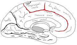 Medial surface of left cerebral hemisphere. | |
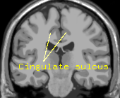 MRI showing the cingulate sulcus in a human. | |
| Details | |
| Identifiers | |
| Latin | sulcus cinguli |
| NeuroNames | 43 |
| NeuroLex ID | birnlex_1468 |
| TA98 | A14.1.09.203 |
| TA2 | 5440 |
| FMA | 83748 |
| Anatomical terms of neuroanatomy [edit on Wikidata] | |
The cingulate sulcus is a sulcus (brain fold) on the cingulate cortex in the medial wall of the cerebral cortex. The frontal and parietal lobes are separated from the cingulate gyrus by the cingulate sulcus. It terminates as the marginal sulcus of the cingulate sulcus. It sends a ramus to separate the paracentral lobule from the frontal gyri, the paracentral sulcus.
Additional images
-
 Position of cingulate sulcus (shown in red).
 -
 Medial surface of right cerebral hemisphere. Cingulate sulcus (labeled as sulcus cinguli) and brain lobes.
 -
 Medial surface of cerebral hemisphere. Medial view. Deep dissection.
 -
 Medial surface of cerebral hemisphere. Medial view. Deep dissection.
 -
 Medial surface of cerebral hemisphere. Medial view. Deep dissection.


External links
Wikimedia Commons has media related to Cingulate sulcus.
- "Anatomy diagram: 13048.000-3". Roche Lexicon - illustrated navigator. Elsevier. Archived from the original on 2012-07-22.
- NIF Search - Cingulate Sulcus via the Neuroscience Information Framework
- v
- t
- e
Anatomy of the cerebral cortex of the human brain
| Superolateral |
| ||||
|---|---|---|---|---|---|
| Medial/inferior |
| ||||
| Both |
| Superolateral | |
|---|---|
| Medial/inferior | |
| Both |
| Superolateral | |
|---|---|
| Medial/inferior |
| Superolateral | |
|---|---|
| Medial/inferior |
|
sulci/fissures
| Superolateral | |
|---|---|
| Medial/inferior |
|
| Parahippocampal gyrus | |
|---|---|
| Cingulate cortex/gyrus | |
| Hippocampal formation | |
| Other |
- Operculum
- Poles of cerebral hemispheres
Some categorizations are approximations, and some Brodmann areas span gyri.
| Authority control databases |
|
|---|
 | This neuroanatomy article is a stub. You can help Wikipedia by expanding it. |
- v
- t
- e


