Cerebellar hemisphere
Each of the two halves of the cerebellum in the brain
| Cerebellar hemisphere | |
|---|---|
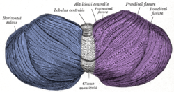 Superior view of the cerebellum. Left cerebellar hemisphere Right cerebellar hemisphere | |
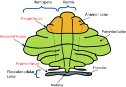 Schematic representation of the major anatomical subdivisions of the cerebellum. Superior view of an "unrolled" cerebellum, placing the vermis in one plane. | |
| Details | |
| Identifiers | |
| Latin | hemisphaerium cerebelli |
| NeuroNames | 1214 |
| NeuroLex ID | birnlex_1575 |
| TA98 | A14.1.07.004 |
| TA2 | 5804 |
| FMA | 76925 |
| Anatomical terms of neuroanatomy [edit on Wikidata] | |
The cerebellum consists of three parts, a median and two lateral, which are continuous with each other, and are substantially the same in structure. The median portion is constricted, and is called the vermis, from its annulated appearance which it owes to the transverse ridges and furrows upon it; the lateral expanded portions are named the hemispheres.
Sections
- The "intermediate hemisphere" is also known as the "spinocerebellum".
- The "lateral hemisphere" is also known as the "pontocerebellum".
- The lateral hemisphere is considered the portion of the cerebellum to develop most recently.[1]
Additional images
-
 Animation.Left cerebellar hemisphereRight cerebellar hemisphere
 -
 Close up animation.
 - Dissection video (45 s). Demonstrating the two cerebellar hemispheres.
-
 Human cerebellum anterior view description (Cerebellar hemisphere is #8)


See also
References
![]() This article incorporates text in the public domain from page 788 of the 20th edition of Gray's Anatomy (1918)
This article incorporates text in the public domain from page 788 of the 20th edition of Gray's Anatomy (1918)
- ^ "Sect. 8, Ch. 6: Functional Subdivisions of the Cerebellum". Archived from the original on 2008-04-01.
External links
Wikimedia Commons has media related to Cerebellar hemisphere.
- Atlas image: n2a3p2 at the University of Michigan Health System
- NIF Search - Cerebellar Hemisphere[permanent dead link] via the Neuroscience Information Framework
- v
- t
- e
Anatomy of the cerebellum
| Lobes | |
|---|---|
| Medial/lateral |
|
| Deep cerebellar nuclei | |
|---|---|
| Cerebellar cortex |
|
| Internal |
|
|---|---|
| Peduncles |
Portal:
 Anatomy

| Authority control databases |
|
|---|
 | This neuroanatomy article is a stub. You can help Wikipedia by expanding it. |
- v
- t
- e


